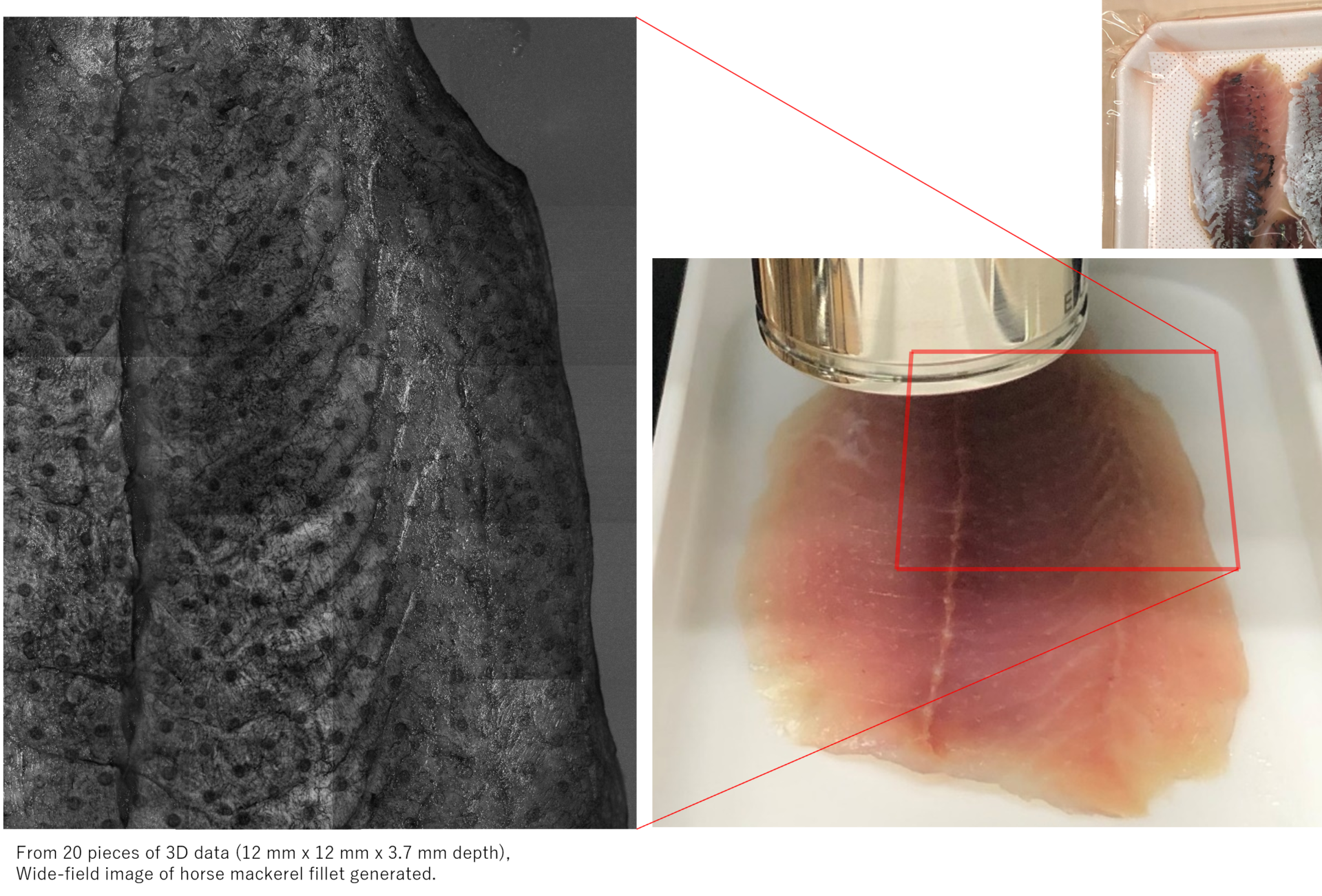
The following is an example of the horse mackerel fillet that was requested by a seafood processing company.
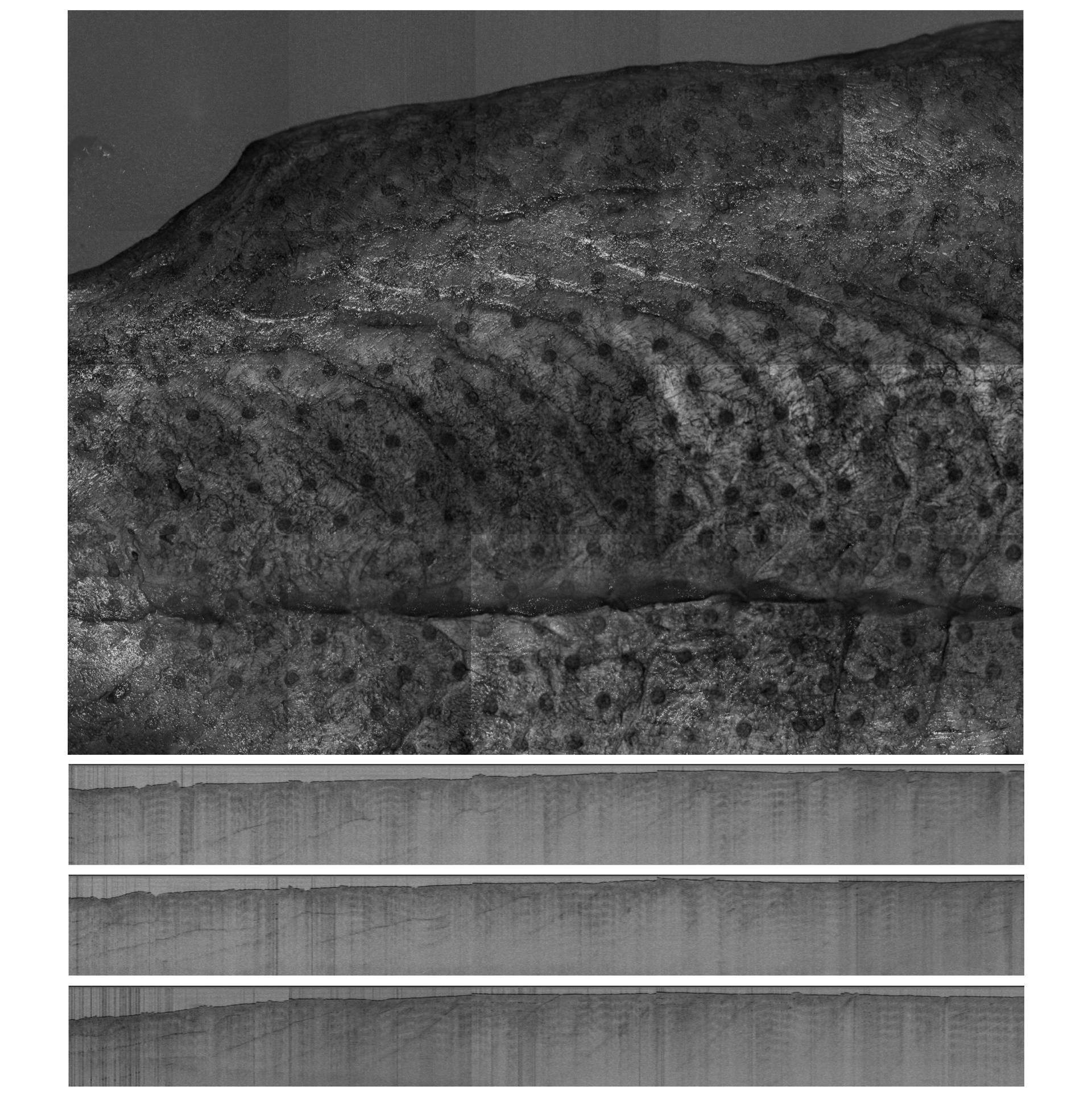
①Projection image
Since the body surface of fish is a protective color (dorsal side = blue or black, ventral side = white), light reflection is too strong on the epidermis side and the signal is disturbed. Therefore, the fillet was measured from the dorsal side.
In the OCT projection image, fine surface morphology was clearly observed.
You can also see the marks (dot pattern) of the round holes made by the water-absorbing sheet. This area appears blackened out in the projected image because the light is scattered in multiple directions, weakening the OCT signal returned from deeper areas.

②Cross-section image
The OCT cross-section image showed the layered structure of the body muscles (orange). The distribution of the intermuscular septum inside the body was also observed (purple).
The round hole marks on the water-absorbing sheet could be distinguished from the others as areas where the surface was rough and uneven (yellow).

The internal structure can be better observed by watching the video.
Cross-section movie of horse mackerel fillet
OCT allows nondestructive measurement of both living and inanimate objects, as long as the probe beam (wavelength 1300 nm) can penetrate deep into the object.
Customization of resolution and measurement speed is also possible.
We will gladly accept your request to try out how it looks. Please feel free to contact us.

